Blood Centrifuge for Clinical and Medical Centrifuged Blood Sample Separations

March 10, 2023 | Centrifuge

Centrifuge for blood samples

Centrifuges are used in clinical applications including the separation of blood samples - a process where the different components of blood are distinguished from one another by weight, size, and density through rotational forces.

Clinical centrifuges come in types according to their application. For instance, a high-speed benchtop model might be used for cell separation; a larger, floor-standing model for high-volume sample processing.

Significance of blood sample centrifuge

Blood samples provide access to an individual’s health and biochemistry. Aside from blood samples, the centrifuge process can also be used to isolate proteins and other molecules from a sample. It can obtain serum or plasma (PRP/Platelet-Rich Plasma centrifuge), which are necessary for assays.

The precise and efficient separation of components allows clinical researchers to identify markers (hormonal levels), gain valuable insight into the underlying biology of medical conditions, and develop effective treatments for them. It also provides an avenue for tracking a disease’s progression and the evaluation of the treatment’s effectiveness over time.

Moreover, this also aids genetic research in the identification of genetic links to specific diseases and conditions. Through analyzing processed blood samples, clinicians can compare individual genetic profiles and determine predispositions. 

Odoo text and image block

Blood Centrifugation: What does a centrifuge do to blood?

In blood component separation, scientists can analyze the target cell population, its purpose, and its function without interference from other cell types. It is a crucial first step in precise diagnosis, leading to improved patient outcomes.

Blood separation centrifuge is commonly used in diagnostic laboratories for screening of disease indicators and in the preparation for transfusions. 

Centrifuging blood samples

Here are the general steps involved in centrifuging blood:

Step 1: Collect a sample of blood. Using a standard venipuncture procedure, collect a small sample of blood. The sample must be collected in a sterile blood tube centrifuge and handled in accordance with the proper sterile technique.

Step 2: Preparing the sample. Place the collected blood sample in a special tube that contains an anticoagulant to prevent the blood from clotting. After securing the seal of the tube, put a label with the necessary information, such as the patient's name and date.

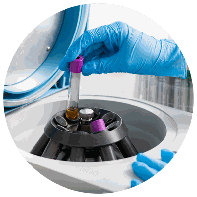
Step 3: Load the sample into the blood centrifuge. Following the manufacturer’s instructions for the specific model, carefully load the sealed blood sample tube into the blood spinner.

Step 4: Setting the speed and time. Setting the speed and time for the centrifugation process should be in accordance with the specific requirements of the separation.

Step 5: Start the blood centrifuge. Start the centrifuge to activate the spinning process.

Step 6: Collect the separated cells. After the centrifugation process is finished, carefully remove the tube from the centrifuge and collect the separated cells. Note that the heavier cells, such as the red blood cells, will be at the bottom of the tube. The lighter cells, such as the platelets, will be at the top.

Step 7: Disposing of the waste. The waste material, along with the anticoagulant and cellular debris, should be properly disposed of according to regulations. 

Other Considerations: 

*Before analysis: separation of white blood cells (WBCs) from red blood cells (RBCs) 

This is done so that analysis is easier for researchers and physicians. There are fewer WBCs than RBCs in the blood. If an initial blood separation phase is not carried out, scientists will have difficulty in acquiring the WBCs for regular clinical testing and research. 

Typically, blood separation procedures produce a distinct layer of white blood cells, known as the Buffy coat, which scientists can remove for further examination.

Blood separation according to the centrifuge’s rotor type

  • Blood separation using a swing-out rotor

       Centrifuges with this type of rotor allow the particles to settle evenly at the tube’s bottom. An advantage       of using this method is the minimal energy consumption. It is also a popular application in medical         laboratories.

  • Blood separation using a fixed-angle rotor

        Centrifuges with a fixed-angle rotor spin the blood components to the opposite side of the tube where            they then slide at the bottom. In conjunction with this type’s increased centrifugal force, the separation         of blood is expedited. This approach is favored by research facilities due to the large speed advantage. 

Odoo text and image block

Recommended blood centrifuge speed and time

  • Time. Researchers found that centrifugation timed 7 or 10 minutes can provide identical test results if compared to the 15-minute time as proposed by the World Health Organization (WHO). This could be a result of improved sample preparation and centrifugation parameters. Thus, shorter centrifugation times could result in (1) more efficient lab operations, (2) reduced errors due to sample contamination and waiting times, and (3) improved test accuracy and patient outcomes. 

  • Speed. The appropriate centrifuge speed is dependent on the intended application of the blood. For diagnostic assays and some research applications, a speed of 4,000 RPM would be adequate; a 6,500 RPM would be right for the majority of research applications. 

Odoo image and text block

Characteristics of a centrifuged blood sample

  • The appearance of three horizontal layers of varied hues. After the tube is removed from the centrifuge, the colored layers can be easily distinguished. Straw-colored plasma layers account for 55% of the total blood volume. The layer beneath the plasma, the Buffy coat, may be white or gray in hue and contains WBCs and platelets. The lower layer is composed of red blood cells, which account for 45% of the total blood volume.

  • The lowest layer of centrifuged blood may be dark red or bright red in color. The color is dependent on the oxygen level of the cells. 

Back To Top
We are fully compliant with the GDPR laws. We promise to safeguard your data and protect your privacy rights.