Typical histopathologic features help in early detection of monkeypox
January 27, 2023 | Histopathology
In the summer of 2022 and amidst an ongoing outbreak, the world became apprehensive of the looming fact that monkeypox (mpox) might just be the next public health emergency. Fear during the early days of COVID was relived as health authorities fumbled for a swift response. With some cases eliciting wide differential diagnosis, avoiding false negatives in order to establish a total traceability of the mpox outbreak is of topmost priority.
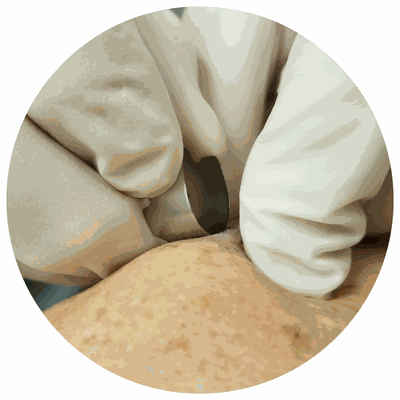
Procedures such as skin biopsy are considered to be of great help. A study done by Dr. Francisco José Rodríguez-Cuadrado, a dermatologist at the Hospital Universitario Puerta de Hierro Majadahondaand, and colleagues identified two of the most defining histopathologic characteristics that could aid in the accurate assessment and early diagnosis of mpox - epidermal necrosis and keratinocytic ballooning. The study, one of the largest of its kind, focused on providing a comprehensive histopathologic and immunohistochemical description of the cutaneous lesions that mpox infection induces.

Outcomes of the study done on 20 male patients with positive mpox virus DNA polymerase chain reaction and immunohistochemial positivity for anti-vaccinia virus, exhibited similar histopathologic findings. According to the researchers, affected areas have thick, central necrosis with irregular hyperplasia and exocytosis of neutrophils with necrotic skin debris. Ballooning on both sides of the necrosis due to pale cytoplasm was also detected while in other cases, swollen pale areas led to intraepidermal vesiculopustules. Advanced lesions showed thick epidermal necrosis consisting of lymphocytes. Conclusively, these cutaneous lesions are strong indicators when it comes to diagnosing mpox.

Researchers divulged that the study’s limitations stem from its limited sample size and the fact that only four cases were studied with an electron microscope.
Awareness among dermatologists and dermatopathologists of the clinical and histopathologic features of the cutaneous lesions of mpox infection plays a massive role in the specific, early diagnosis and subsequent adequate treatment.